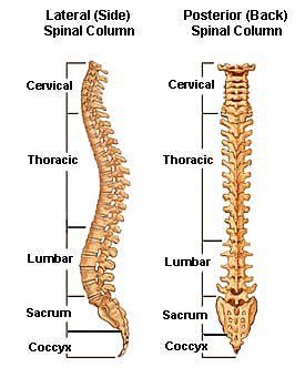
Neck/Cervical spine has 7 vertebrae, C1 – C7
Chest/Thoracic spine has 12 vertebrae, T1 – T12
Low Back/Lumbar spine has 5 vertebrae, L1 – L5
Pelvis/Sacrum spine has 5 fused vertebrae S1 – S5
Tail bone/Coccyx has 3 vertebrae
The Cervical Spine
The cervical spine is further divided into two parts; the upper cervical region (C1 and C2), and the lower cervical region (C3 through C7). C1 is termed the Atlas and C2 the Axis. The Occiput (CO), also known as the Occipital Bone, is a flat bone that forms the back of the head.

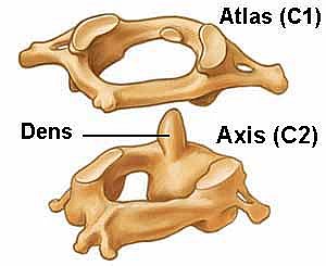
Atlas (C1)
The Atlas is the first cervical vertebra and therefore abbreviated C1. This vertebra supports the skull. Its appearance is different from the other spinal vertebrae. The atlas is a ring of bone made up of two lateral masses joined at the front and back by the anterior arch and the posterior arch.
Axis (C2)
The Axis is the second cervical vertebra or C2. It is a blunt tooth–like process that projects upward. It is also referred to as the ‘dens’ (Latin for ‘tooth’) or odontoid process. The dens provides a type of pivot and collar allowing the head and atlas to rotate around the dens.
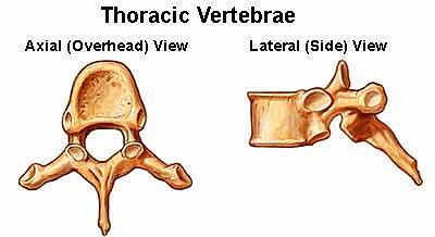
Thoracic Vertebrae (T1 – T12)
The thoracic vertebrae increase in size from T1 through T12. They are characterized by small pedicles, long spinous processes, and relatively large intervertebral foramen (neural passageways), which result in less incidence of nerve compression.
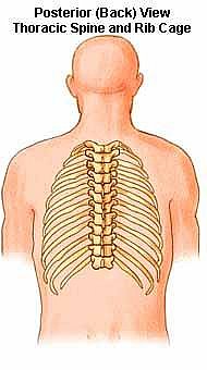
Ribs
The rib cage is joined to the thoracic vertebrae. At T11 and T12, the ribs do not attach and are so are called “floating ribs.” The thoracic spine’s range of motion is limited due to the many rib/vertebrae connections and the long spinous processes.
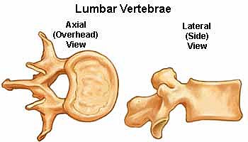
Lumbar Vertebrae (L1 – L5)
The lumbar vertebrae graduate in size from L1 through L5. These vertebrae bear much of the body’s weight and related biomechanical stress. The pedicles are longer and wider than those in the thoracic spine. The spinous processes are horizontal and more squared in shape. The intervertebral foramen (neural passageways) are relatively large but nerve root compression is more common than in the thoracic spine.
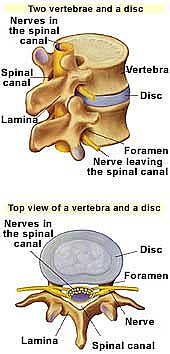
Intervertebral Discs
The bony vertebrae of your spinal column are separated from one another by “pads” of tough cartilage called intervertebral discs. These discs act like “shock absorbers” during activity, allowing the spine to move freely. When these curves are in their normal alignment, your body is in a balanced position. This spreads your weight evenly throughout the vertebrae and discs, so you are less likely to have strain and injury. The center of each intervertebral disc is made up of a gelatin-like substance surrounded by a fiber-like outer lining. As your body ages, the disc’s nucleus begins to stiffen. This reduces the flexibility and increase the chances that discs may “rupture,” especially in the lumbar spine which carries so much of the body’s weight.
The spinal cord, which begins at the base of the brain and runs within the spinal canal, ends in the lumbar spine area in a bundle of nerves known as the cauda equina. The spinal canal runs through the center of the spinal column and protects the spinal cord and other delicate spinal nerves.
At each vertebrae level, a pair of nerves branch off from the spinal cord or the cauda equina (one to the left and one to the right). These spinal nerve roots are part of the body’s “electrical” system, carrying a “current” (for sensation and movement) to specific parts of the body. If the disc gets damaged, due to it’s proximity to the nerve roots, the disc may bulge and impinge a nerve and transmission may be affected.
Learn how Chiropractic works
Click Here